What’s the difference between cardiac MRI and cardiac CT
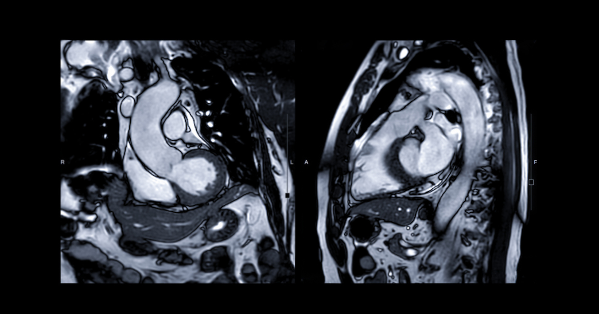

Over the past decade, Cardiac Magnetic Resonance Imaging (CMRI) and Cardiac Computed Tomography (CCT) have become indispensable tools for cardiac imaging because of their ability to generate extraordinarily vivid images of the beating heart and blood vessels in a strictly non-invasive manner.
According to Samer Sayyed, Cardiovascular Disease specialist at Nebraska Medicine, there are five characteristic differences between cardiac MRI and CT:
Radiation and Safety
Cardiac MRI utilizes magnetic fields in combination with radio waves and powerful computers to generate images. Cardiac CT on the other hand relies on shooting X-Rays from multiple angles to produce a full three-dimensional computer model of the body and internal organs. The radiation involved with CT may result in an increased lifetime risk of cancer; especially in people who’ve had repeated CT scans. Cardiac MRI is radiation free.
Need for Intravenous Contrast
An intravenous injection of contrast media is absolutely necessary for cardiac CT to define the blood-tissue interface and tell the difference between different kinds of tissues. While with cardiac MRI, IV contrast isn’t needed, there are some occasions when a doctor might need to use contrast during a cardiac MRI to highlight conditions that would be otherwise be undetectable.
Contrast Side Effects
There are two types of contrast used: iodine-based (for cardiac CT) and gadolinium-based (for cardiac MRI). The iodine-based agents used for CT have the potential to cause “Contrast Induced Nephropathy” in some patients. That is a condition that can cause reduction in kidney function for patients with pre-existing kidney problems. Gadolinium-based contrast agents, on the other hand, are much safer for the kidneys but have the potential to cause a condition known as “Nephrogenic Systemic Fibrosis” in those with reduced kidney function. Ultimately, iodine-based agents can be removed during hemodialysis if needed whereas the fate of gadolinium based agents is less clear.
Heart Rate Control
To produce blur-free images of the beating heart, both CT and MRI require a fairly regular heart rate. Ideally, that is 60 beats per minute with a CT scan. While detrimental to cardiac CT, rapid heart rates are usually not much of an issue for cardiac MRI as long as the pulse remains regular.
Patient-scanner Compatibility
Because cardiac MRI uses strong magnets, a very thorough screening for ferromagnetic objects on or within the patient’s body needs to be conducted before any patient is deemed safe to undergo cardiac MRI. Fortunately, patients with implanted ferromagnetic devices can safely undergo cardiac CT provided they have good kidney function and are not severely allergic to iodinated contrast media.


