What is capsule endoscopy, and why is it used?

If a patient is experiencing chronic diarrhea, abdominal pain, unexplained weight loss or bleeding, a capsule endoscopy, or video capsule endoscopy, may provide answers. A capsule endoscopy is a non-invasive diagnostic procedure used to examine the gastrointestinal tract.
Nebraska Medicine gastroenterologist Ishfaq Bhat, MD, tells us what capsule endoscopy is used for and how it differs from other methods.
How does capsule endoscopy work?
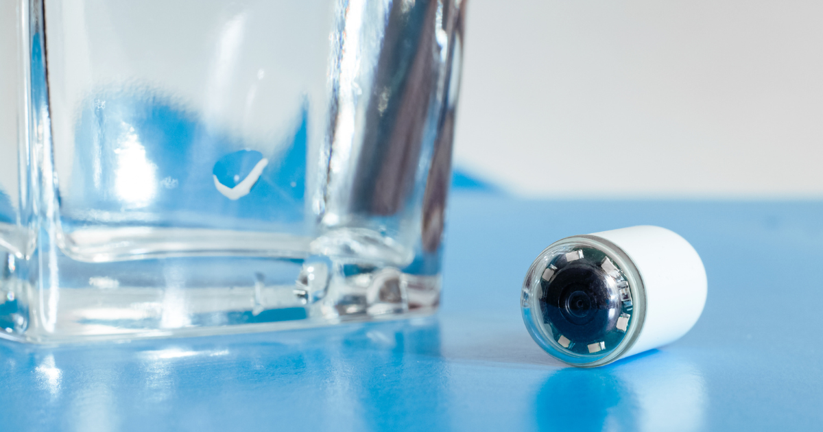
“Video capsule endoscopy is a wireless, noninvasive diagnostic procedure that uses a small, pill-sized camera to capture images of the GI tract, particularly the small intestine,” says Dr. Bhat. “The patient swallows the capsule, wears a belt recorder and goes home with appropriate instructions.”
The video capsule is about the size of a large vitamin pill and passes safely through the digestive system. Multiple images are taken per second and transmitted wirelessly to the belt recorder.
The light source on the capsule illuminates the GI tract, allowing for clear images. The patient returns the recorder the following day, and images are downloaded to a proprietary software. A gastroenterologist reviews the images on a computer frame by frame. These images can identify inflammation, ulcers, bleeding, tumors, polyps or signs of Crohn’s or celiac disease.
"Capsule endoscopy is very good at detecting problems in the small intestine, especially for issues like unexplained bleeding and other conditions, with a success rate of 60% to 80%. However, its accuracy can be reduced if the capsule moves too quickly through the intestines or if there’s difficulty visualizing due to food or other material in the bowel," says Dr. Bhat.
What does capsule endoscopy diagnose?
Capsule endoscopy is commonly used to diagnose conditions such as:
- Small bowel bleeding (occult or overt) – To identify sources of bleeding in the small intestine that are not visible with other imaging methods.
- Crohn’s disease – An inflammatory bowel disease that affects the intestines.
- Celiac disease – An autoimmune disorder where ingestion of gluten harms the small intestine.
- Polyps or ulcers – To identify small growths or sores in the intestines.
- GI tumors or cancer – Detecting growths or abnormalities in the small intestine or GI tract.
- Malabsorption disorders – Conditions where the intestines can’t absorb nutrients.
How does capsule endoscopy differ from other methods?
Capsule endoscopy is considered noninvasive and more comfortable compared to other diagnostic methods for examining the GI tract. Traditional endoscopy, such as an upper endoscopy or colonoscopy, involves inserting a flexible tube with a camera through the mouth or rectum.
“With capsule endoscopy, there’s no need for sedation or scope insertion,” says Dr. Bhat. “In most patients, it can examine the entire small bowel. Unlike CT or MRI, capsules provide live, real-time images of the lumen of the small bowel.”
While capsule endoscopy provides clear imaging of the small intestine, it doesn’t allow for therapeutic interventions such as biopsy or removing polyps during the procedure.
Who should get a capsule endoscopy?
Good candidates for capsule endoscopy include:
- Patients with unexplained GI bleeding or anemia.
- Those with suspected bowel disorders (e.g., Crohn’s disease).
- Individuals who can’t tolerate conventional endoscopy.


